
INCISORS REPLACEMENT: CASE REPORT
- lyly mong
- Dec 22, 2024
- 1 min read

A female patient, aged 50, was referred for treatment. She perviously had two incisors #11 and #21 of PFM crown and metal post core fabricated for more than 5 years ago. Pre-operative x-ray shows periapical lesions on the related teeth, with asymptomatic periapical abscess.


Figure 1.
PFM crown and metal post-core was removed, and root canal treatment was performed. The coronal tooth structure provides adequate ferrule for post core and crown on endontically treated tooth.


Figure 2.
Using Shining 3D Intra-oral scanners.


Figure 3.
Cementation of zirconia post core with G-Cem Cement.


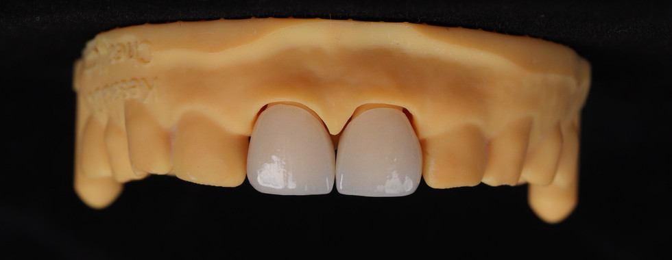

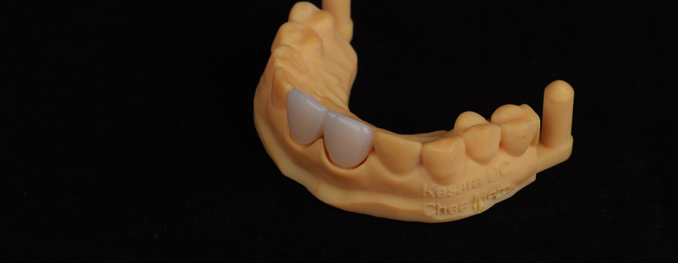
Figure 4.
Digital Impressions with intraoral scans. 3D print model was printed and zirconium crowns was fabricated.

Here is the Final Result after Cementing
highlights video will be added later … thanks for watching


Comments